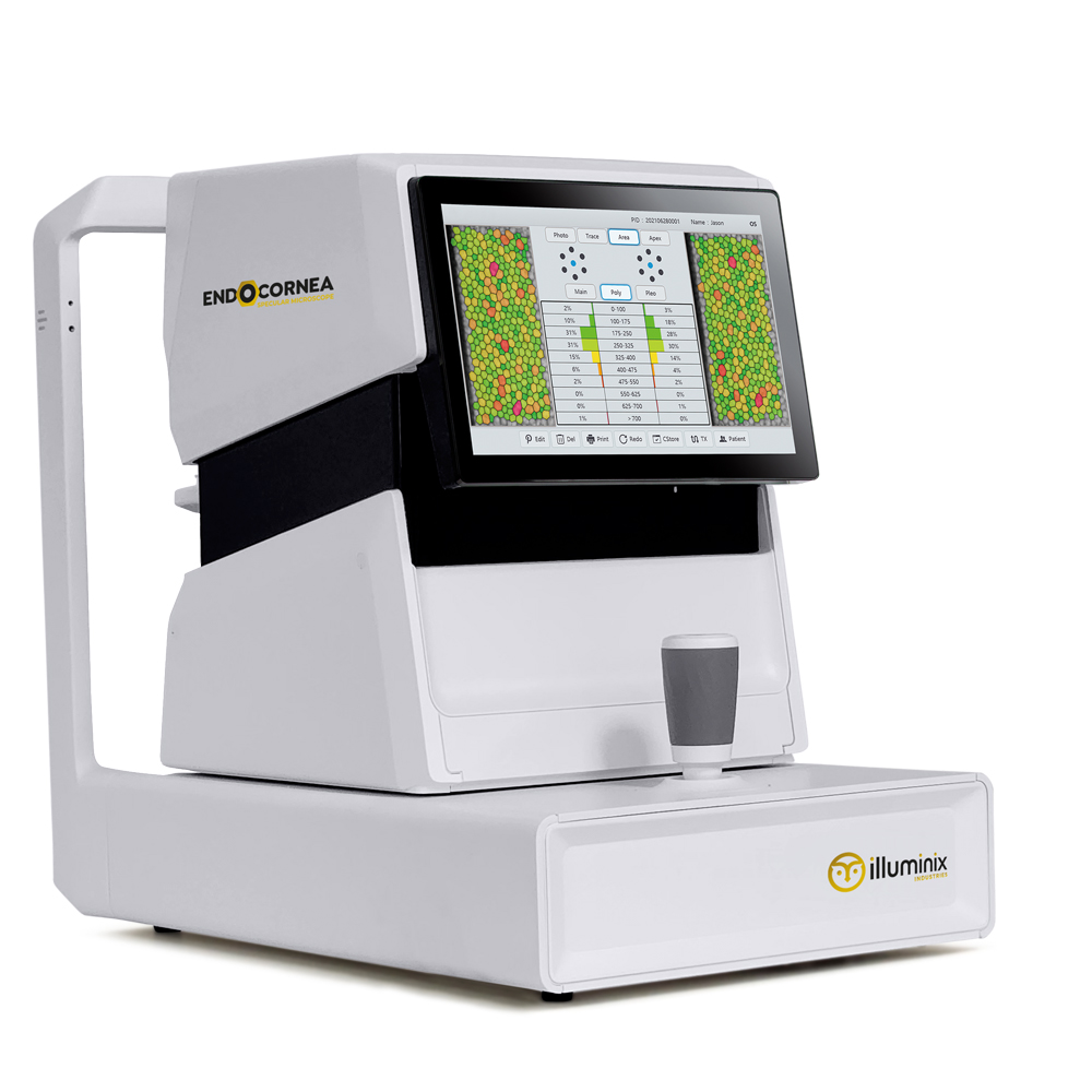
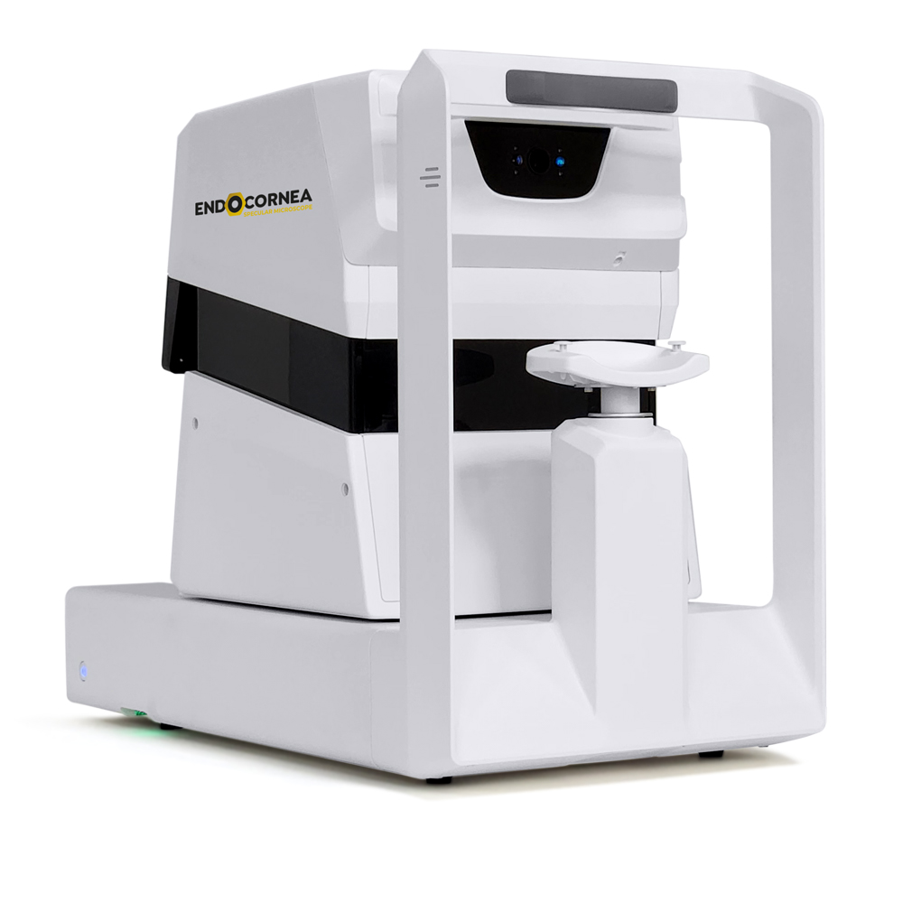
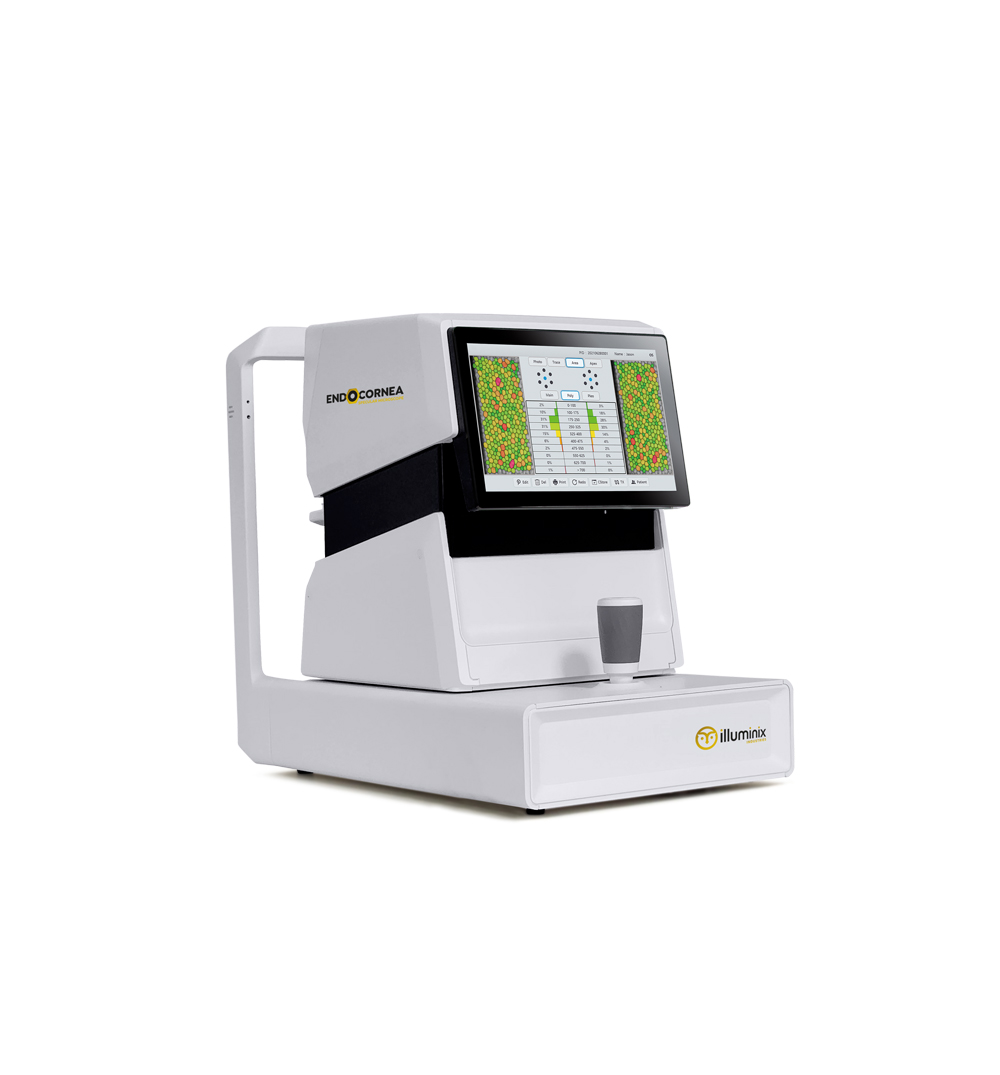
CORNEAL SPECULAR MICROSCOPE
Fully automatic
Fast measurement and analysis
Fully automatic working mode:
Automatic pupil detection, automatic alignment, automatic shooting, automatic analysis.
Concise user interface, one-click operation experience.
Wide field of view & multiple measuring points
0.25mm x 0.55mm wide field endothelial cell imaging.
There are as many as 13 measuring points, including: central measuring point, 6 paraxial measuring points and 6 edge measuring points.
Large-capacity database
Built-in high-performance main frame.
Up to 1TB storage capacity.
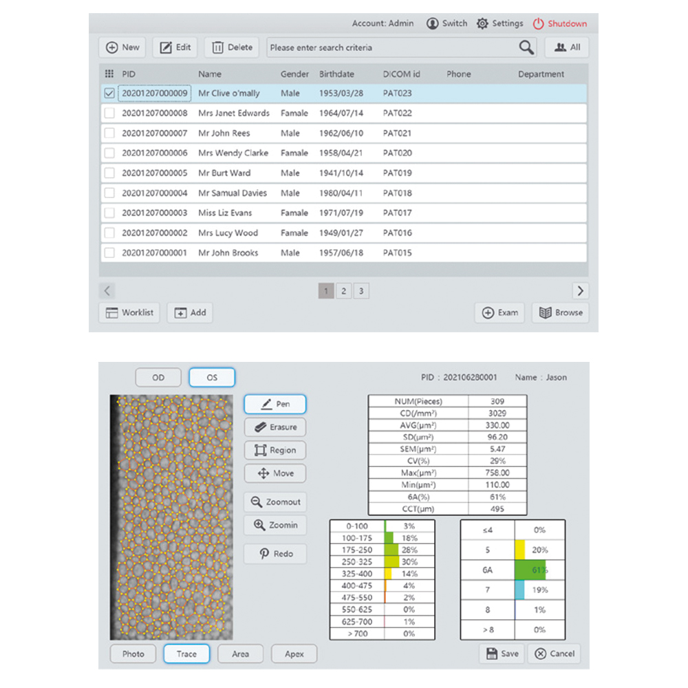
Multi functional case management system allows convenient and efficient operation of medical records.
Specification:
Capturing method: non-contact
Capturing range: 0.25mm x 0.55mm
Capturing area: Center and 12 peripheral fixation points
Working mode: Full-auto/Semi-auto
Cornea thickness measurement range: 400μm ~ 750μm
Cornea thickness measurement accuracy:
±10μm (<600μm); ±25μm (> 600μm)
Analysis parameter: NUM (Number of cells)
CD (Cell density)
AVG (Average area)
SD (Standard deviation of area)
CV (Coefficient of Variation)
MAX (Maximum area)
MIN (Minimum area)
6A (Percentage of hexagonal cells)
Histogram: Area: Polymegathism (Distribution by areas)
Apex: Plemorphism (Distribution by shapes)
Display: 10.4” colored touchscreen(1080P)
Printing: Able to connect to most regular printing devices
Data output: USB x 2; LAN x 1; support DICOM 3.0
Dimensions and weight: 315 x 535 x 465mm; approx. 23Kg
Power source: 100V AC ~ 240V AC; 50/60Hz; 100VA


